5 Tips for Mastering Cell Organelles Coloring

Embarking on the colorful journey of mastering cell organelles can be both an educational and entertaining endeavor. Coloring is not just a fun activity but also an effective way to learn about the intricate architecture of cells. In this blog, we will delve into five comprehensive tips to enhance your understanding of cell organelles through the art of coloring.
1. Understand the Basics

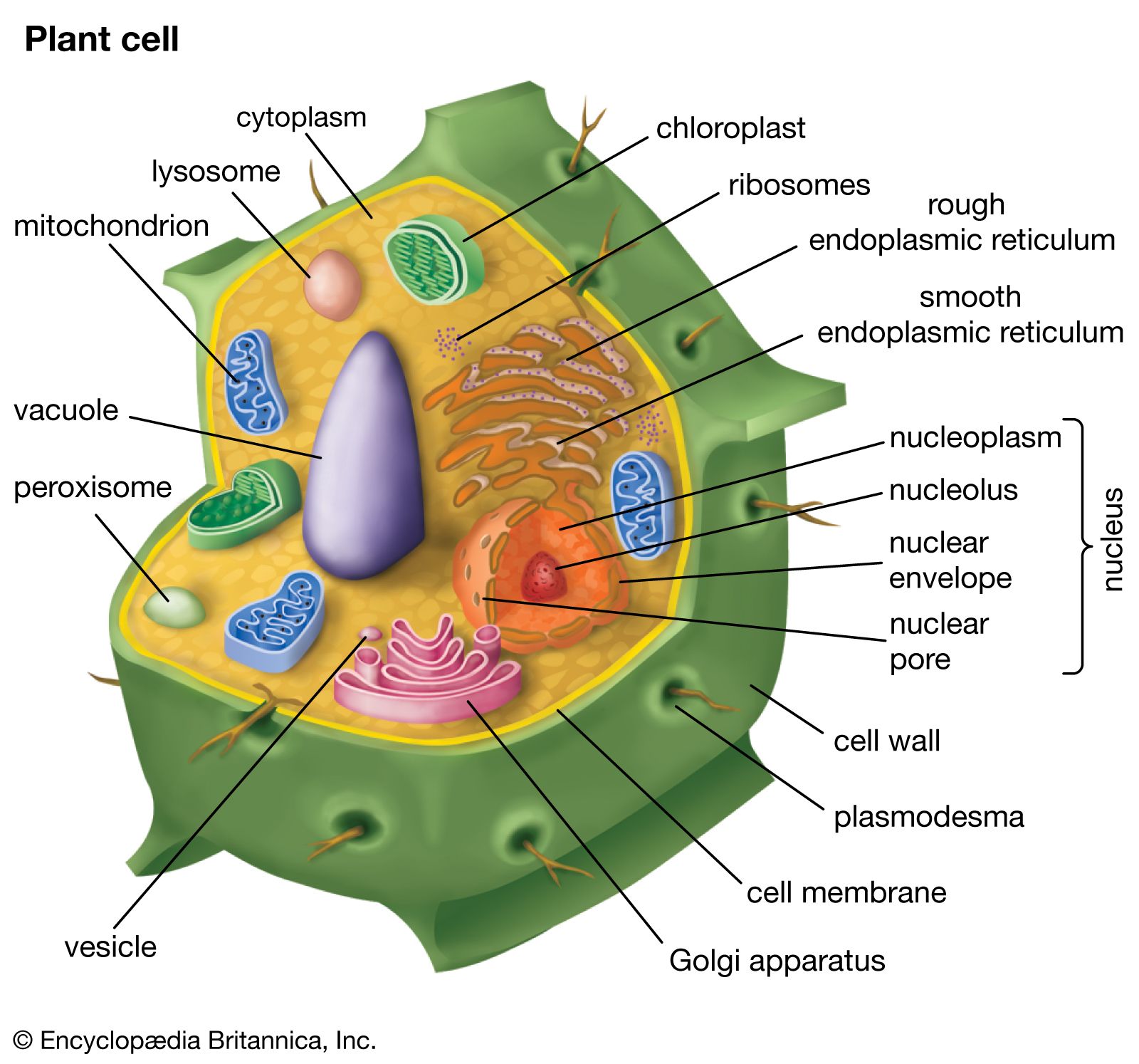
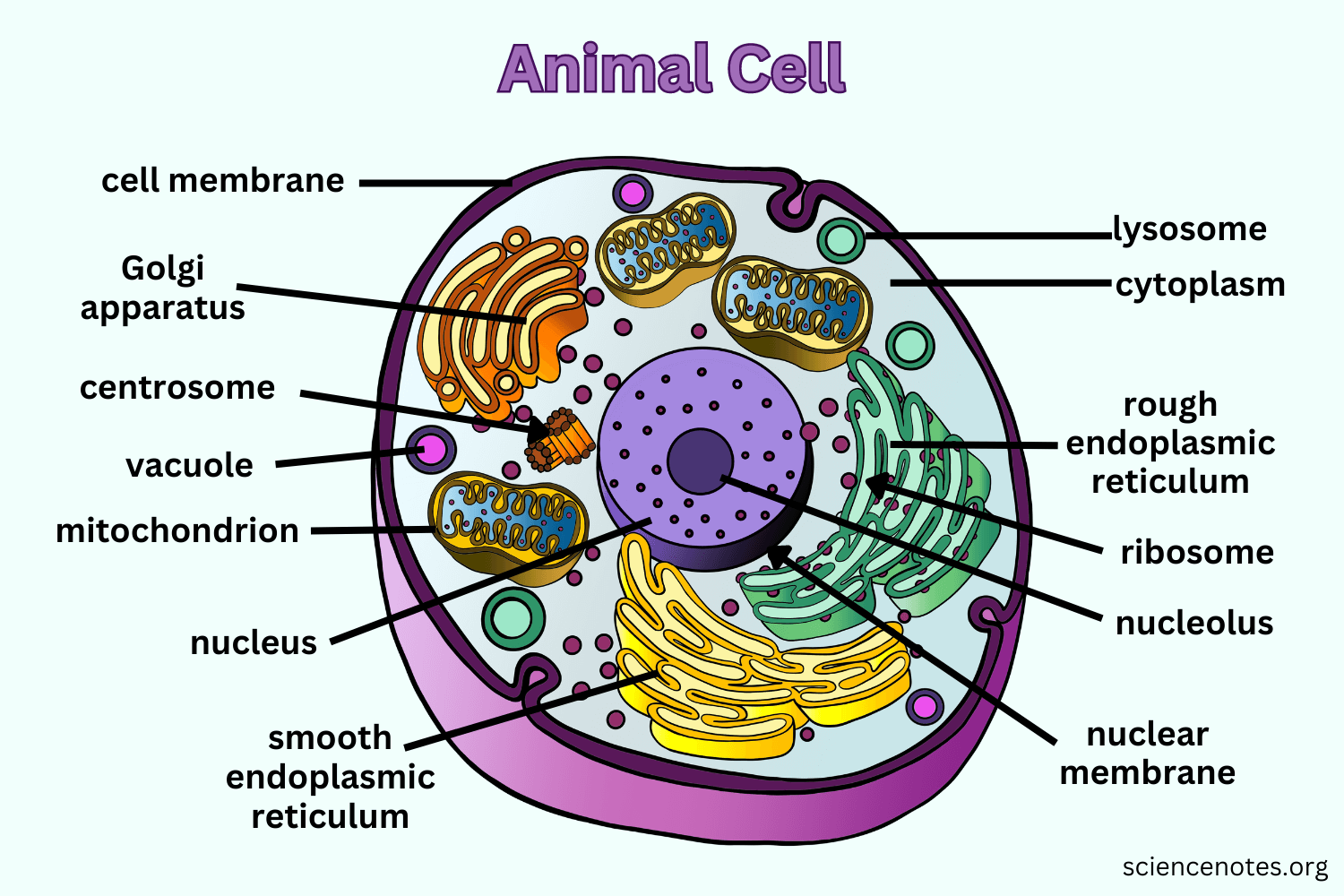
Before diving into your coloring palette, it’s crucial to understand the basics of cell structure. Cells are the fundamental units of life, and organelles are their functional compartments, each performing specific tasks. Here’s a quick rundown:
- Nucleus: The control center of the cell, housing genetic material.
- Mitochondria: Known as the powerhouse, where energy production occurs.
- Endoplasmic Reticulum: A network responsible for protein synthesis (Rough ER) and lipid metabolism (Smooth ER).
- Golgi Apparatus: Packages and modifies proteins for transport.
- Lysosomes: Digests waste materials and cellular debris.
- Chloroplasts: Exclusive to plant cells, where photosynthesis takes place.
- Vacuole: Storage for water, nutrients, and waste.
📚 Note: Getting familiar with cell organelles before coloring can make the process more meaningful and efficient.
2. Use a Color Key

A color key is your roadmap to success when coloring cell organelles. Assigning each organelle a specific color not only helps in immediate identification but also in retaining information. Here’s how you can create one:
| Organelle | Color |
|---|---|
| Nucleus | Purple |
| Mitochondria | Orange |
| Endoplasmic Reticulum (Rough) | Blue |
| Endoplasmic Reticulum (Smooth) | Light Blue |
| Golgi Apparatus | Green |
| Lysosomes | Red |
| Chloroplast | Green (with black outline for grana) |
| Vacuole | Yellow |

Keep this key handy as you color. Consistency in color selection helps in reinforcing the association between the organelle and its function.
3. Practice Visualization Techniques

Visualization is key to internalizing the 3D nature of cells. Here are some techniques to practice:
- Use Perspective: Try to depict the depth of cell organelles. Overlap structures to show how organelles might look in a real cell.
- Shade with Light: Simulate light falling on the organelles to give them dimension. This not only looks realistic but also aids in understanding their spatial arrangement.
- Integrate Textures: Different textures can indicate different organelle functions. For instance, ribosomes on the Rough ER can be shown as small dots or bubbles.
Remember, the goal is to get a feel for the spatial arrangement within the cell, enhancing your mental model of cellular biology.
4. Incorporate Real-life Applications

Biology doesn’t exist in isolation. Connecting the anatomy of cells to real-life applications can solidify your understanding:
- Medical Science: How lysosomal storage diseases relate to lysosome function.
- Environmental Impact: The role of chloroplasts in photosynthesis and how this affects global ecosystems.
- Biotechnology: The use of organelles like mitochondria for energy production in genetically modified organisms.
This contextual learning approach not only broadens your knowledge base but also enriches your coloring experience by making it more relatable.
5. Make it Interactive

Interactive learning through coloring can be exceptionally engaging. Here are ways to make your sessions more dynamic:
- Label as You Go: Place labels directly on the cell diagram to visually connect the organelle to its name and function.
- Create Stories: Invent narratives around the organelles, like mitochondria being the cell’s energetic workforce.
- Use Apps or Digital Tools: Color organelles on a tablet or computer where you can easily change colors, undo mistakes, or simulate different lighting effects.
- Collaborative Coloring: Work with peers on a large-scale cell model, discussing each organelle’s role as you go.
Engaging with the material in this way turns passive learning into an active, memorable experience.
By integrating these tips into your cell organelles coloring sessions, you not only enhance your understanding of cellular biology but also make the process enjoyable and memorable. With a color key, visualization techniques, real-world applications, and interactive methods, you're on your way to mastering cell biology. Each stroke of color adds not just to your artwork but also to your knowledge, making learning an art form in itself.
To conclude, mastering the nuances of cell organelles through coloring involves a blend of science and art. It's a journey where understanding the cell's structure aids in your coloring endeavor, and your creative approach to coloring deepens your comprehension of cell biology. Enjoy the process, let your creativity flow, and watch as your knowledge of cellular biology takes on a vibrant life of its own.
Why should I use different colors for each organelle?

+
Using different colors for each organelle facilitates visual memory. When you associate specific functions with colors, it becomes easier to remember and recognize organelles in diagrams or actual cell images.
How does coloring cell organelles help with studying?

+
Coloring cell organelles engages multiple senses, making the learning experience more vivid and memorable. It also reinforces the structure and function of cells through visual and kinesthetic learning.
Can I use digital tools for coloring cell organelles?

+
Absolutely! Digital coloring tools can offer features like easy color correction, shading, and even 3D representations of cells. This can be particularly useful for students looking for more interactive learning experiences.


