5 Key Facts About Skeletal Muscle Anatomy Worksheet

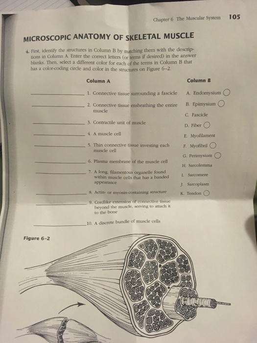
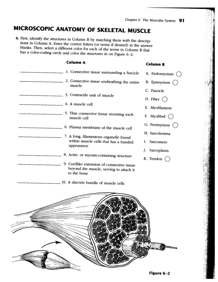
Understanding the anatomy of skeletal muscles is crucial for students in various fields, from physical therapy to sports science. To help learners grasp this essential topic, we'll delve into 5 Key Facts About Skeletal Muscle Anatomy using a structured worksheet approach. This guide will break down complex anatomical details into easily digestible parts, enhancing understanding and retention.
Fact 1: Muscle Fiber Structure

Skeletal muscles are composed of muscle fibers, which are long, cylindrical cells. Here’s how to understand the structure:
- Myofibrils: These are elongated, thread-like structures inside the muscle fiber that are responsible for muscle contraction.
- Sarcomeres: Myofibrils consist of repeating units called sarcomeres, the contractile unit of a muscle fiber. Each sarcomere has:
- Actin Filaments: Thin filaments where cross-bridges can form.
- Myosin Filaments: Thick filaments that contain myosin heads for attaching to actin.
- Z-discs: They mark the boundaries of each sarcomere.
- Other Components:
- Mitochondria for energy production.
- Sarcoplasmic Reticulum for calcium storage, essential for muscle contraction.
✅ Note: When studying muscle fiber structure, remember that muscle contraction occurs when the actin and myosin filaments slide past each other, reducing the length of the sarcomere.
Fact 2: Muscle Contraction Mechanism

Muscle contraction involves several intricate steps:
- Neuromuscular Junction Stimulation: A nerve impulse releases acetylcholine at the neuromuscular junction, which depolarizes the sarcolemma.
- Calcium Release: The depolarization triggers the release of calcium ions from the sarcoplasmic reticulum.
- Cross-Bridge Cycling: Calcium binds to troponin on the actin filament, causing a conformational change that exposes binding sites for myosin heads.
- Power Stroke: Myosin heads attach to actin, pivot, and then release, pulling the actin towards the center of the sarcomere.
- Relaxation: When nerve impulses cease, calcium pumps back into the sarcoplasmic reticulum, stopping the cross-bridge activity.
⚠️ Note: Muscle relaxation is as crucial as contraction to prevent muscle spasms and ensure proper movement.
Fact 3: Muscle Types

There are three main types of skeletal muscle fibers based on their contraction speed and fatigue resistance:
- Slow Oxidative Fibers (Type I):
- High in myoglobin, giving them a red color.
- Excellent endurance, low force, and slow to fatigue.
- Fast Glycolytic Fibers (Type IIB):
- White in color, due to low myoglobin.
- Fast, high force, and quick to fatigue.
- Fast Oxidative-Glycolytic Fibers (Type IIA):
- Pink in color, intermediate properties.
- Moderate speed, force, and fatigue resistance.
💡 Note: An individual's muscle composition can be genetically influenced but can also adapt to training and lifestyle.
Fact 4: Muscle Attachments

Muscles attach to bones via:
- Tendons: Connect muscle to bone, allowing for efficient movement.
- Aponeuroses: Broad, sheet-like tendons that attach muscles to bones or other muscles.
- Origins: The attachment point where the muscle is relatively immobile or has less movement.
- Insertions: The point where the muscle moves the bone.
📌 Note: Understanding origins and insertions helps predict movement based on muscle contraction.
Fact 5: Muscle Shapes

Muscles can be categorized by their shape, influencing their function:
- Fusiform: Biceps brachii, spindle-shaped, good for muscle force concentration.
- Parallel: Rectus abdominis, fibers run parallel, allowing for long movement and high force.
- Pennate:
- Unipennate: Oblique muscles, with muscle fibers attaching at an angle on one side.
- Bipennate: Rectus femoris, fibers attach on both sides.
- Multipennate: Deltoid, fibers attach to multiple tendons from multiple angles.
- Triangular: Pectoralis major, fan or triangular shape, for broad movements.
| Muscle Shape | Example |
|---|---|
| Fusiform | Biceps Brachii |
| Parallel | Rectus Abdominis |
| Unipennate | Extensor Digitorum |
| Bipennate | Rectus Femoris |
| Multipennate | Deltoid |

In summary, understanding skeletal muscle anatomy is vital for a comprehensive grasp of human movement, exercise physiology, and rehabilitation. Each fact mentioned provides insight into the complexity of muscles, from their microscopic structure to their macroscopic organization, highlighting how muscles are perfectly designed for function. This worksheet approach not only simplifies the topic for learners but also ensures retention through the structured presentation of key facts.
Why is it important to understand muscle fiber types?

+
Understanding muscle fiber types helps tailor training programs to enhance specific performance attributes like endurance or power.
How do muscles and tendons work together?

+
Muscles generate force, while tendons transmit this force to the bone, ensuring movement.
Can muscle shape affect functionality?

+
Yes, different muscle shapes optimize the muscle’s ability to generate force and adapt to specific movement patterns.


