Cell Diagram Worksheet: Master the Basics Simply

Understanding cell diagrams is a foundational step for anyone studying biology, be it at high school or university level. A cell diagram not only outlines the structural components of cells but also elucidates how these components work together to sustain life. In this comprehensive guide, we'll delve into the intricacies of cell diagrams, highlighting key elements and offering tips for mastering them.
Why Understanding Cell Diagrams is Crucial

Cell diagrams are vital for:
- Visualization: They provide a visual representation of complex cellular structures, making it easier to comprehend their form and function.
- Education: They aid in the teaching of cell biology by simplifying abstract concepts through graphical depiction.
- Research: Scientists use these diagrams to explain findings or hypothesize new functions within the cell.

Key Components of a Cell Diagram
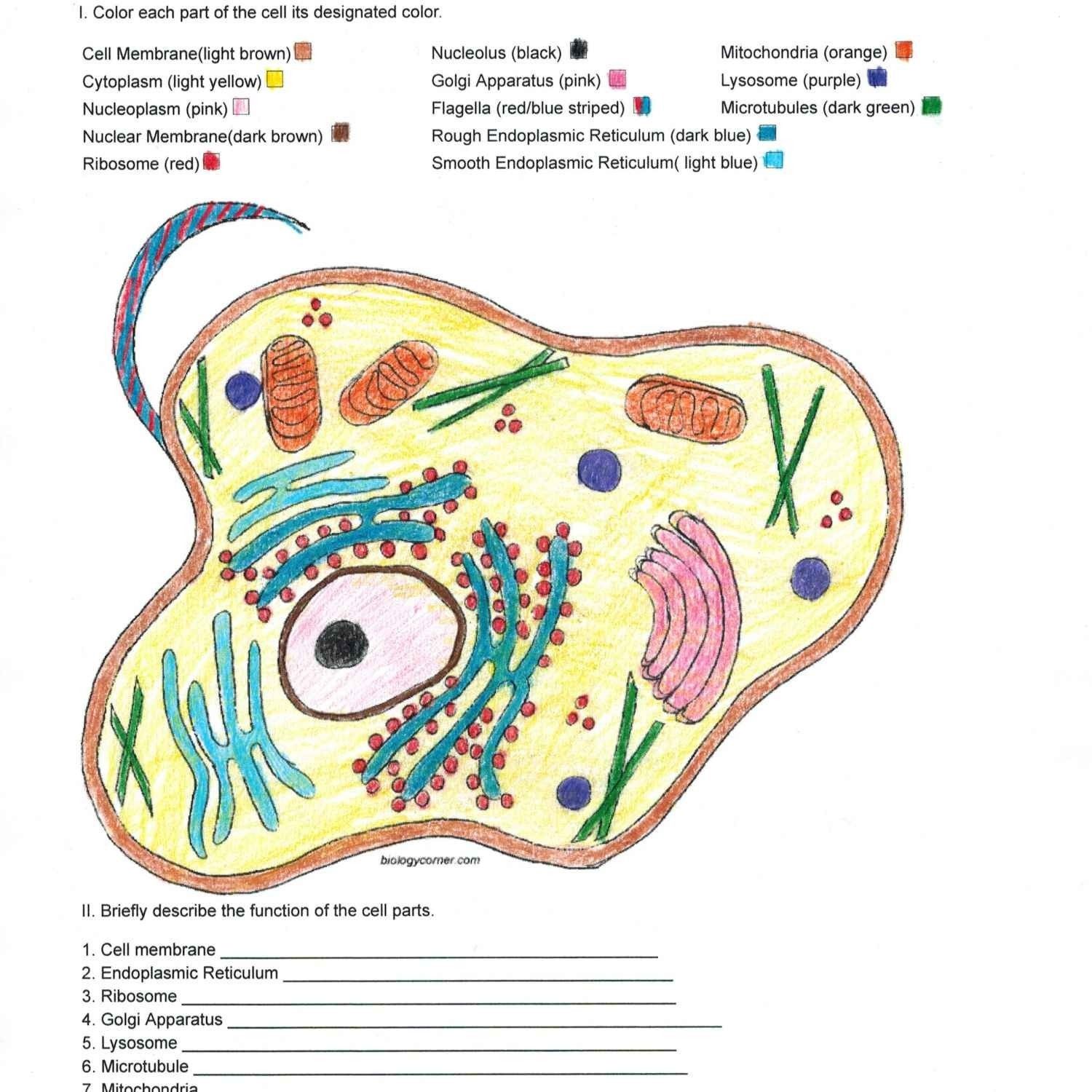
Here are the essential parts typically depicted in cell diagrams:
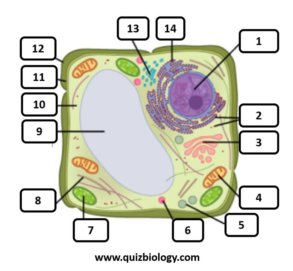
- Plasma Membrane: The outermost layer that regulates what enters and leaves the cell.
- Cytoplasm: A jelly-like substance where most of the cell's metabolic activities occur.
- Nucleus: Contains the cell's DNA; it controls all cellular functions.
- Endoplasmic Reticulum: Involved in protein synthesis (rough ER) and lipid synthesis (smooth ER).
- Golgi Apparatus: Modifies, sorts, and packages proteins and lipids for secretion or use within the cell.
- Mitochondria: Known as the powerhouse of the cell, it generates ATP through cellular respiration.
- Ribosomes: Sites of protein synthesis.
- Lysosomes: Digest cellular waste and foreign substances.
- Vesicles: Small membrane-bound sacs used for transport inside the cell.
- Cytoskeleton: Provides structure and allows for movement within the cell.

How to Study Cell Diagrams Effectively
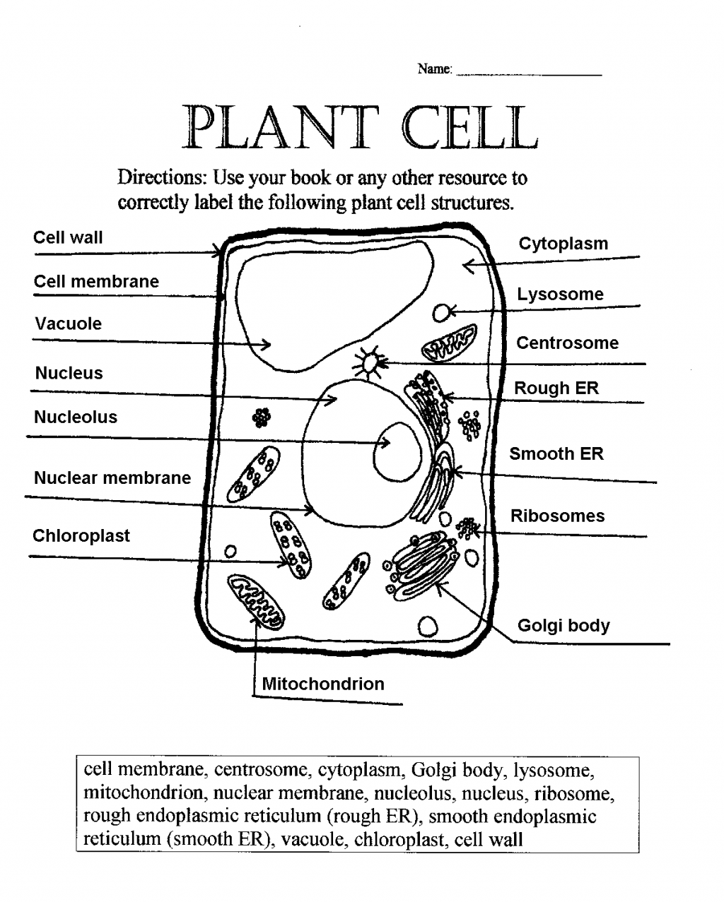
Here are strategies to master cell diagrams:
- Labeling: Practice labeling diagrams from memory. Use flashcards or interactive tools online.
- Comparative Analysis: Compare diagrams of different cell types to understand structural and functional differences.
- Create Your Own: Draw diagrams to reinforce your understanding. Start with simple sketches and gradually add detail.
- Color Coding: Use colors to differentiate between organelles, which helps in visual memory retention.
- Active Learning: Engage with diagrams through questions, quizzes, or by teaching someone else.
🎨 Note: When drawing your own cell diagrams, focus on accuracy over complexity. Simple, correctly labeled diagrams are more beneficial for learning than overly detailed ones.
Common Mistakes in Cell Diagrams

Here are some frequent errors to avoid:
- Incorrect Labeling: Ensure all labels are correct; wrong labels lead to misconceptions.
- Scale Discrepancy: Cells have different sizes; keep the scale in mind when drawing or interpreting diagrams.
- Omission of Structures: Don’t skip any parts, even if they are small or less common in all cells.
- Misunderstanding Function: Remember, diagrams show structure, but understanding the function is equally important.
Tips for Teachers and Students
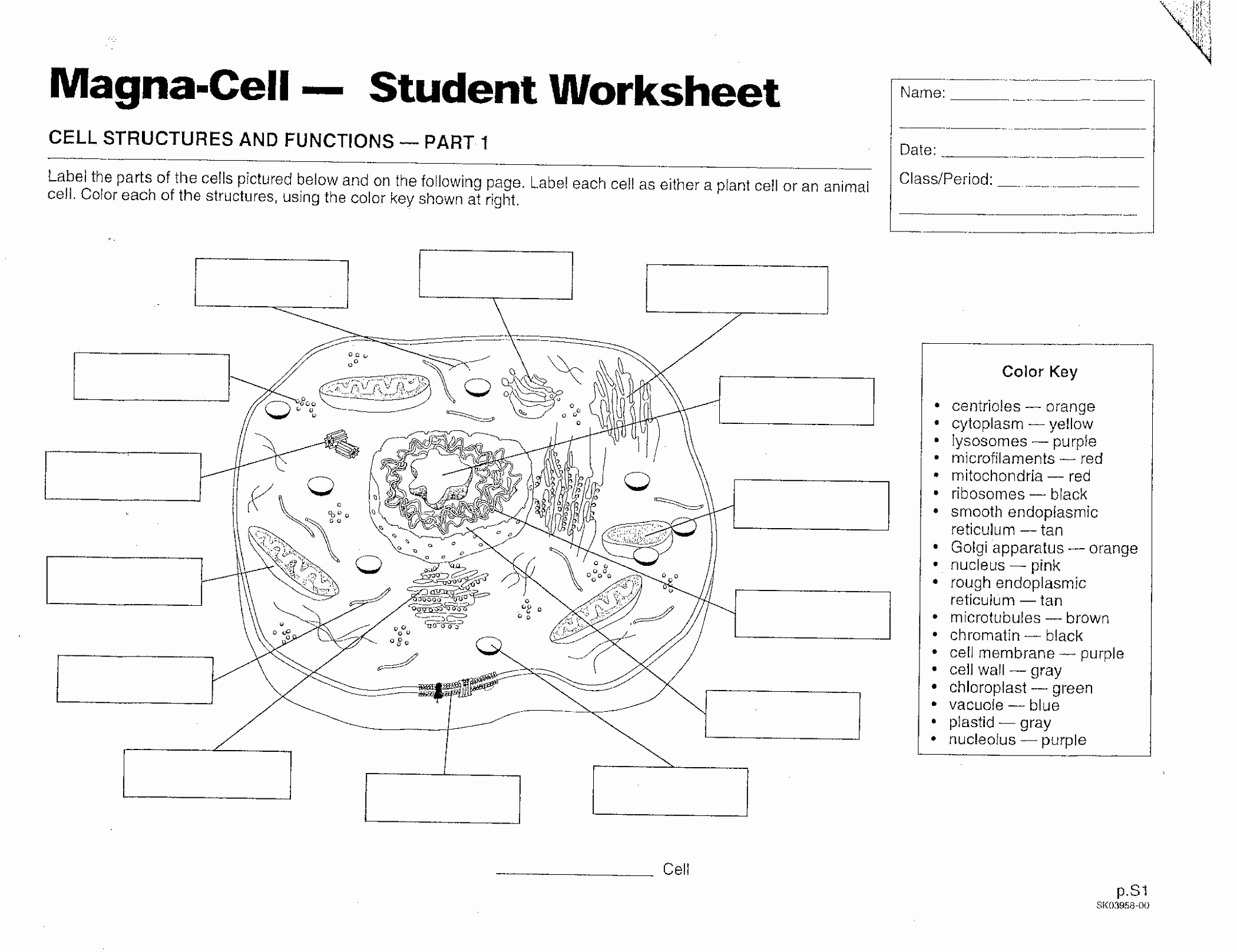
Here are insights for both teaching and learning from cell diagrams:
- Dynamic Learning: Use animations or video tutorials to understand the dynamic processes within cells.
- Interactive Diagrams: Utilize software or apps that allow for interaction with cell parts.
- Analogy Use: Compare cellular components to real-life objects or systems for better retention.
- Group Work: Engage in group projects to discuss and clarify misunderstandings.
- Regular Revision: Keep revisiting cell diagrams periodically to commit them to long-term memory.
In our journey through the intricate world of cell diagrams, we've covered the why and how of learning about cellular structures. These diagrams not only serve as a visual guide but also as a tool for deeper understanding. By mastering the basics of cell diagrams, students can unlock the secrets of life itself, understand complex biological processes, and excel in their studies. Whether through active learning, drawing, or group discussions, the path to cell diagram mastery is filled with discovery and learning.
What are the differences between animal and plant cell diagrams?
+
Plant cell diagrams include structures like the cell wall, chloroplasts, and a large central vacuole, which are absent in animal cells. Additionally, animal cells often have lysosomes and centrioles, which plant cells do not.
Can you explain the function of ribosomes?

+
Ribosomes are the sites of protein synthesis within cells. They translate the genetic code from mRNA into chains of amino acids, which fold into proteins.
Why is the Golgi Apparatus significant in cell function?
+
The Golgi Apparatus modifies, sorts, and packages proteins and lipids from the endoplasmic reticulum. It ensures that these molecules are either secreted from the cell or correctly placed within it for use.
