Sheep Heart Dissection Worksheet: Explore Anatomy with Ease
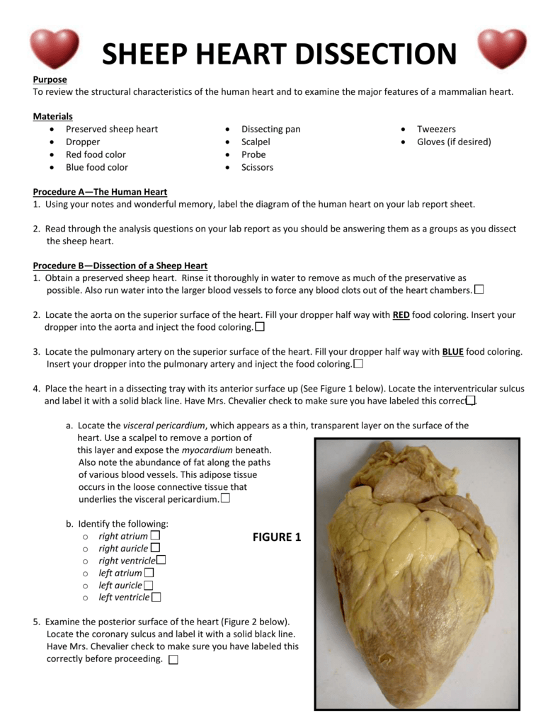
Heart dissection offers an intriguing journey into the internal workings of this vital organ, crucial for understanding biological systems, especially for those pursuing studies in biology, medicine, or veterinary sciences. By exploring the anatomy of a sheep's heart, you gain hands-on experience that enhances your comprehension of cardiac structure and function. This worksheet provides a detailed guide for those who want to delve into the fascinating world of cardiac dissection, making it accessible for students at all levels.
Why Dissect a Sheep’s Heart?
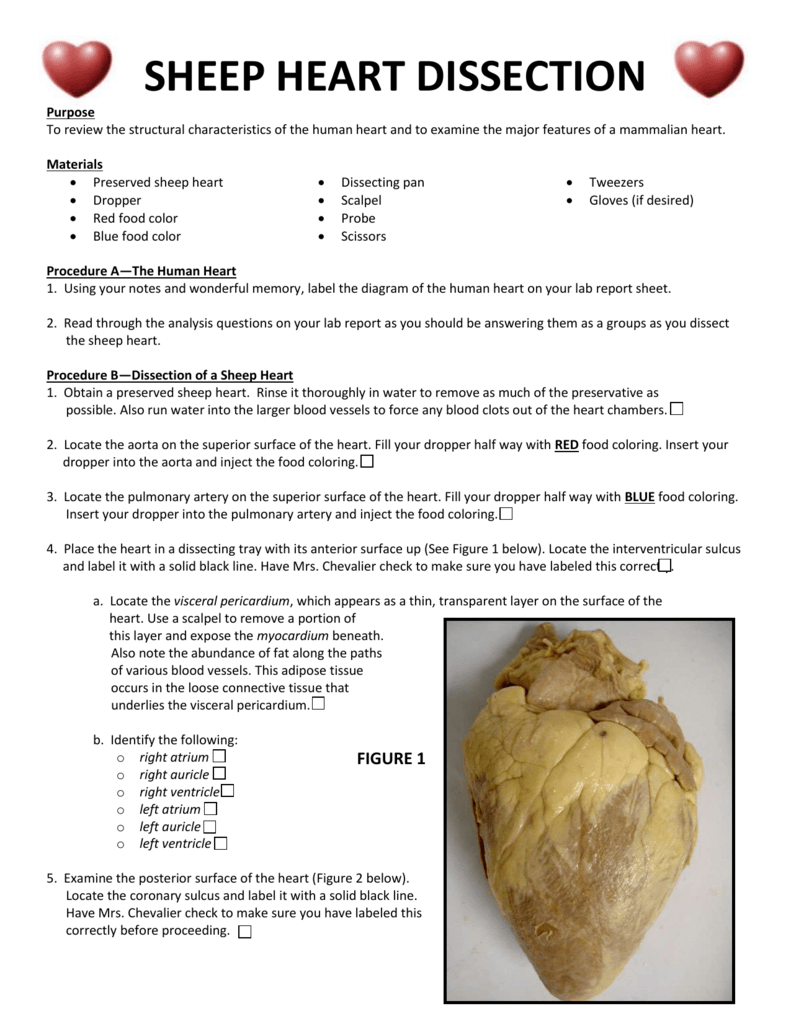
The sheep heart is an excellent choice for educational purposes due to several reasons:
- Similarity to the Human Heart: Sheep hearts have similar anatomical structures to human hearts, providing a good basis for comparison and study.
- Availability: Sheep hearts are easily sourced from slaughterhouses, making them readily available for educational use.
- Size: The size of a sheep heart is large enough to make internal structures easily visible for observation.
📚 Note: Always ensure that the use of animal parts for educational purposes complies with your institution’s ethical guidelines and local regulations.
Preparation for Dissection
Materials Needed
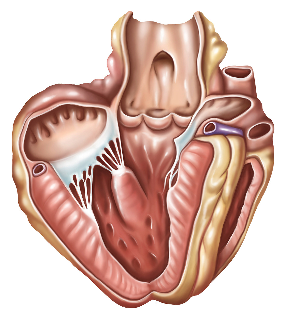
- A sheep heart preserved in formalin or freshly obtained.
- Dissection tools: Scalpel, scissors, forceps, pins, and a dissecting probe.
- Dissection tray or a plastic container.
- Gloves for protection.
- Safety goggles to shield your eyes from splashes.
- Lab coat or apron for cleanliness.
Preparation Steps
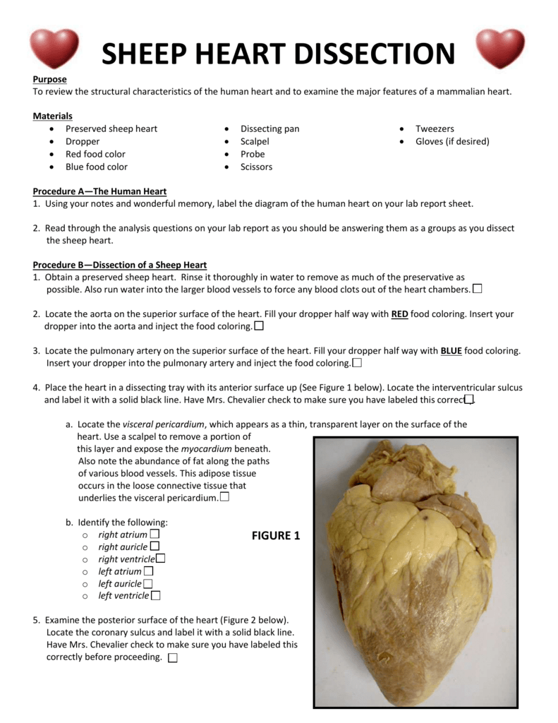
- Wash your hands thoroughly before starting.
- Put on your protective gear, including gloves, goggles, and a lab coat.
- Place the sheep heart in the dissection tray, ensuring it is securely positioned for easy handling.
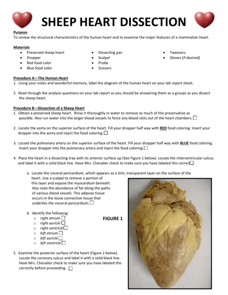
- Take a moment to observe the external features of the heart:
- Note the apex and the base of the heart.
- Examine the blood vessels, particularly the aorta, pulmonary artery, and vena cavae.
- Identify the pericardium, the double-walled sac that encloses the heart, if it’s intact.
Dissection Process
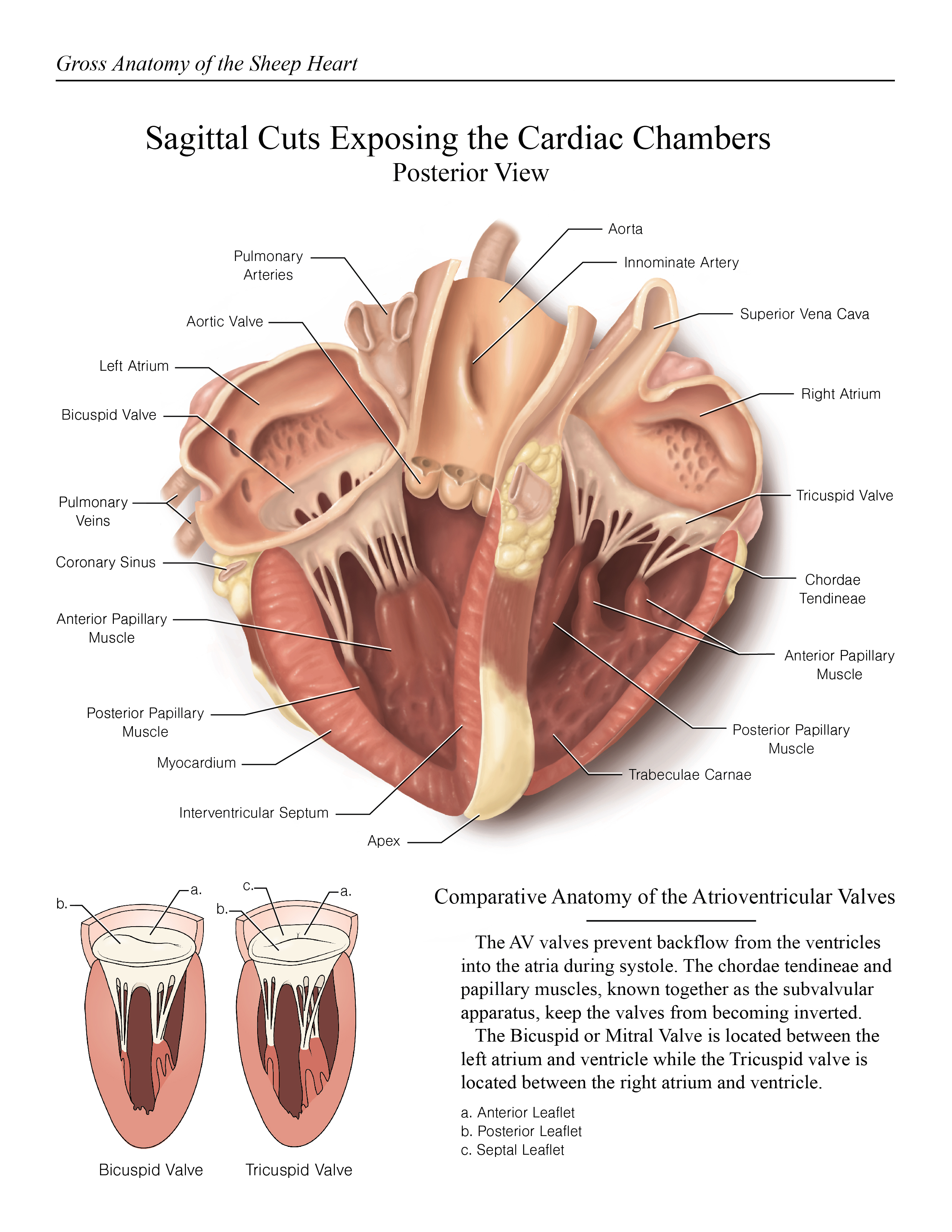
The following steps guide you through the dissection process:
External Features

- Start by examining the pericardial sac, which, if present, should be gently removed to reveal the heart muscle beneath.
- Locate the atria and ventricles.
- The atria are on the top, often identifiable by their thinner walls, while ventricles are more muscular at the bottom.
- Find the coronary vessels supplying the heart with blood.
Internal Features

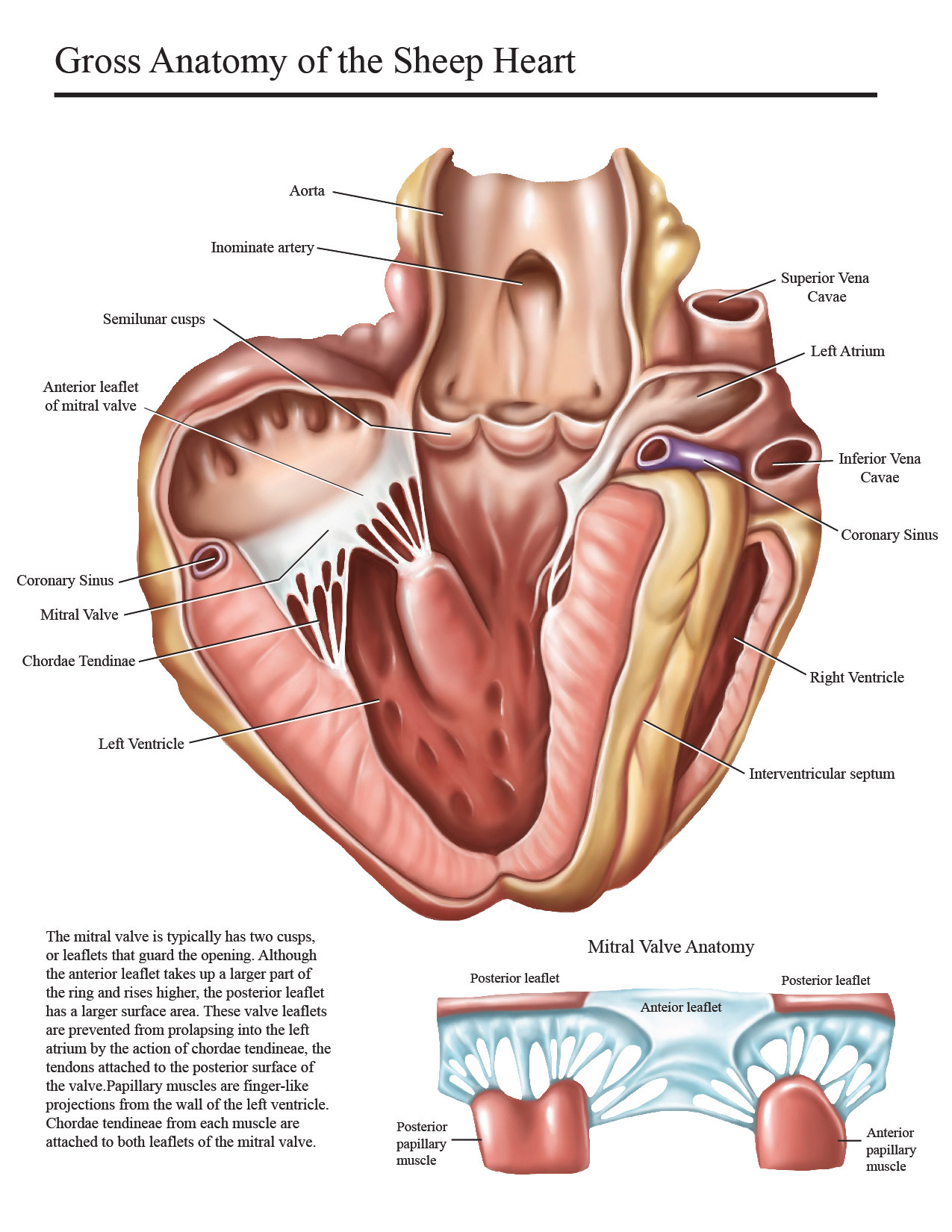
- Make a longitudinal cut through the right ventricle, extending up to the right atrium.
🔍 Note: Be careful to cut just deep enough to expose the interior without damaging the valves.
- Identify the:
- tricuspid valve, which separates the right atrium from the right ventricle.
- chordae tendineae and papillary muscles that support the valve.
- The septum that divides the right and left halves of the heart.
- Proceed to cut through the left ventricle and left atrium:
- The left side is much more muscular due to the higher pressure needed to pump blood to the body.
🔬 Note: Compare the thickness of the muscular walls between the left and right ventricles.
- Identify the:
- mitral valve or bicuspid valve, between the left atrium and ventricle.
- Look for the aortic and pulmonary valves at the base of their respective arteries.
Further Exploration

Examine the Great Vessels

- Identify the aorta and trace its path from the heart.
- Follow the pulmonary artery, noting its branch to the lungs.
- Locate the vena cavae, superior and inferior, which return blood to the heart.
Study the Blood Flow Path
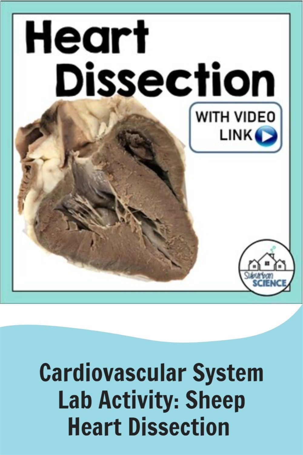
| Chamber/Vessel | Description | Blood Flow |
|---|---|---|
| Right Atrium | Receives deoxygenated blood | From superior and inferior vena cava |
| Right Ventricle | Pumps blood into pulmonary circulation | Through pulmonary valve to lungs |
| Left Atrium | Receives oxygenated blood | From pulmonary veins |
| Left Ventricle | Pumps blood into systemic circulation | Through aortic valve to body |
Disposal and Clean-Up

After the dissection, ensure proper disposal:
- Dispose of the dissected heart and gloves in biohazardous waste containers.
- Clean all tools and the dissection tray thoroughly with a disinfectant.
- Wash your hands and clean any surfaces that might have come into contact with the specimen.
Wrapping Up the Experience

This journey through the anatomy of a sheep’s heart not only provides a practical understanding of cardiac structure but also instills a deeper appreciation for cardiovascular functions. By engaging with the dissection process, you’ve observed the intricate design that allows our hearts to pump life-sustaining blood throughout our bodies. Remember, this experience can be enhanced by revisiting the anatomy through textbooks or online resources, and discussing your observations with peers to further consolidate your learning. Each step in this guide not only brings you closer to understanding the heart but also equips you with the skills for future anatomical studies.
Why is it important to dissect a sheep’s heart for educational purposes?

+
Dissecting a sheep’s heart provides hands-on learning, allowing students to observe and understand the intricate anatomy and physiology of the heart, which is similar to the human heart, thereby enhancing their comprehension of complex biological systems.
What are some safety precautions to take during heart dissection?

+
Ensure you wear protective gear like gloves, safety goggles, and a lab coat or apron. Dispose of biological materials in designated biohazard containers, clean tools and surfaces with disinfectant, and maintain a hygienic environment throughout the process.
Can the heart dissection be performed without affecting the valves?

+
Yes, with careful cutting, you can avoid damaging the valves. It’s crucial to make gentle, precise cuts to expose the chambers while preserving the delicate structures of the heart, like the valves.
