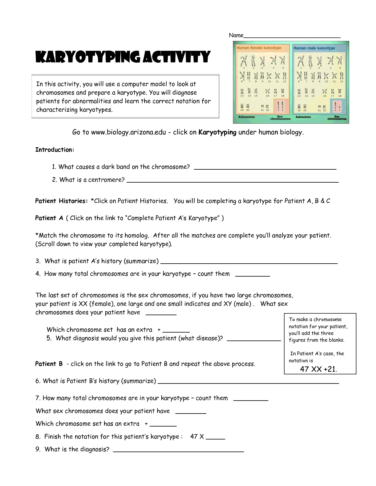
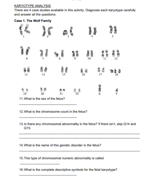
Karyotyping Activity: Chromosomal Adventure Awaits in this Worksheet!

Embarking on a journey through the intricate world of human genetics, karyotyping provides an educational and exploratory avenue into our biological blueprint. This worksheet introduces students, educators, and biology enthusiasts to the fascinating process of identifying and analyzing chromosomes in a cell, referred to as karyotyping. Our chromosomal adventure not only unveils the structure and characteristics of human chromosomes but also emphasizes their significance in understanding genetic diseases and the broader landscape of human evolution.
Why Karyotyping Matters

Karyotyping isn't just a scientific technique; it's a window into our genetic past and future. This method allows us to:
- Diagnose chromosomal abnormalities leading to genetic disorders.
- Identify chromosomal sex.
- Track the inheritance of chromosomal traits.
- Assist in personalized medicine and genetic counseling.
Getting Started with Your Karyotyping Worksheet


Before diving into the process, familiarize yourself with the necessary tools and materials:
- Microscope slides
- Stains (Giemsa or other chromosome-specific stains)
- Photographs or high-resolution images of chromosomes
- Scissors and glue for hands-on arrangement
- Chromosome image sets for matching and pairing
Step-by-Step Karyotyping Process

Follow these steps to perform karyotyping:
- Cell Collection: Cells are harvested from an individual, often from blood or amniotic fluid.
- Cell Culture and Stimulation: The cells are cultured to stimulate cell division.
- Metaphase Arrest: Colcemid is added to halt cell division at metaphase.
- Cell Lysis: The cells are treated with a hypotonic solution, which causes them to swell, spreading the chromosomes.
- Chromosome Staining: Stain the chromosomes using Giemsa or a similar stain to reveal banding patterns.
- Slide Preparation: Spread the chromosomes onto a slide for microscopic examination.
- Photography and Capture: Use a photomicrograph or digital imaging to capture the chromosomes.
- Chromosome Arrangement: Manually or digitally arrange the chromosomes by size, shape, and banding pattern.
- Karyotype Analysis: Examine the karyotype for any abnormalities or genetic anomalies.
📝 Note: Karyotyping can detect numerical and structural chromosome abnormalities, but it does not reveal single-gene mutations.
Identifying Chromosomes and Their Bands

Each chromosome has a unique banding pattern that can be used for identification:
- Chromatids: The two identical copies of DNA in a duplicated chromosome.
- Centromere: The constricted region where chromatids are joined.
- Arms (p and q): The short (p) and long (q) arms of the chromosome.
- Bands: Alternating light and dark stripes created by staining, indicating specific segments.
| Chromosome Number | Group | Key Characteristics |
|---|---|---|
| 1-3 | A | Large metacentric chromosomes |
| 4-5 | B | Submetacentric |
| 6-12, X | C | Medium-sized, metacentric or submetacentric |
| 13-15 | D | Acrocentric with satellites |
| 16-18 | E | Small metacentric or submetacentric |
| 19-20 | F | Small metacentric |
| 21-22, Y | G | Small acrocentric, the Y being the smallest |

The Importance of Chromosome Abnormalities

Abnormal karyotypes can lead to various genetic conditions:
- Down Syndrome (Trisomy 21): An extra chromosome 21.
- Turner Syndrome (Monosomy X): Missing an X chromosome in females.
- Klinefelter Syndrome (XXY): An extra X chromosome in males.
- Cri-du-chat Syndrome: Deletion on the short arm of chromosome 5.
⚠️ Note: While karyotyping is useful, conditions like cystic fibrosis or Huntington's disease, which result from single gene mutations, are not detectable through this method.
Advancing Beyond Traditional Karyotyping

Modern techniques have advanced karyotyping:
- Fluorescence In Situ Hybridization (FISH): Highlights specific DNA sequences with fluorescent probes.
- Comparative Genomic Hybridization (CGH): Identifies copy number variations in the genome.
- Chromosomal Microarray Analysis (CMA): Provides high-resolution maps of chromosomal imbalances.
Recap of Our Chromosomal Adventure

Through this worksheet, we have delved into the intricate world of karyotyping, a pivotal technique in the realm of genetics. From collecting cells to analyzing their chromosomal content, we've explored how this method can reveal the human genetic story. By understanding the structure, function, and potential disorders associated with our chromosomes, we gain insight not only into the basic principles of genetics but also into the practical applications that impact our health, genetic counseling, and even legal decisions in paternity cases or forensic science.
What is karyotyping used for?

+
Karyotyping is used to diagnose chromosomal abnormalities, determine genetic sex, investigate stillbirths or miscarriages, provide genetic counseling, and in cancer diagnosis to assess chromosomal changes in tumors.
Can karyotyping detect all genetic disorders?

+
No, karyotyping can detect chromosomal abnormalities but cannot identify single-gene mutations or small deletions/duplications that are responsible for some genetic disorders.
How is karyotyping performed?

+
Karyotyping involves collecting cells, stimulating cell division, arresting it at metaphase, lysing cells to spread chromosomes, staining, photographing, arranging, and analyzing the chromosomes for structural and numerical abnormalities.


