Heart Dissection Lab Worksheet Answers Revealed

If you're studying biology, particularly anatomy and physiology, you'll likely encounter labs where you get hands-on experience with dissections. One of the most common dissections in high school and college biology courses is the dissection of a mammalian heart. This experience not only provides tangible insight into the cardiovascular system but also teaches valuable dissection techniques. In this comprehensive blog post, we'll dive into the answers and insights from a heart dissection lab worksheet, enhancing your understanding of the heart's structure and function.
Introduction to Heart Dissection

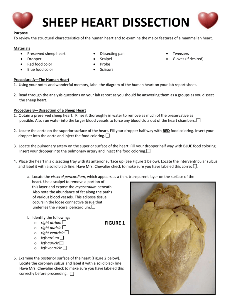
Heart dissection is often an eye-opening experience for students. It provides a 3D perspective of the heart, allowing one to see the chambers, the flow of blood, and the textures of heart tissues in real life, which diagrams and models can’t fully replicate. Here are some initial steps and considerations:
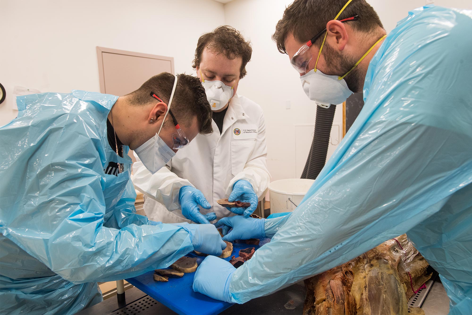
- Ensure you have the necessary tools: scalpels, scissors, tweezers, pins, and gloves.
- Study the external anatomy first before cutting.
- Understand the heart’s orientation: the apex points down, and the base is broader, facing upwards.
Understanding the Heart’s Structure

Let’s explore what you should look for during the dissection:
External Anatomy

Before making any incisions:
- Identify the Apex and Base of the heart.
- Locate the major blood vessels: the Aorta, Pulmonary Trunk, Vena Cavae (superior and inferior).
- Observe the atria’s auricles, which are ear-shaped extensions.
Internal Anatomy

Upon opening the heart:
- Left and Right Atria - At the top, separated by the interatrial septum.
- Left and Right Ventricles - The more muscular chambers, with the left being the thickest due to its role in systemic circulation.
- Interventricular Septum - The muscular wall separating the two ventricles.
- Valves:
- Atrioventricular (AV) Valves: Tricuspid on the right, Mitral or Bicuspid on the left.
- Semilunar Valves: Pulmonary Valve and Aortic Valve.
Flow of Blood Through the Heart

Understanding blood flow through the heart can be challenging, but here’s how it goes:
- Deoxygenated blood enters the right atrium via the vena cavae.
- It passes through the tricuspid valve into the right ventricle.
- From the right ventricle, it exits through the pulmonary trunk to the lungs via the pulmonary arteries for oxygenation.
- Oxygenated blood returns via pulmonary veins into the left atrium.
- It then goes through the mitral valve into the left ventricle.
- Lastly, it exits the heart through the aortic valve into the aorta, distributing oxygenated blood to the body.
Common Questions During Dissection

Here are some common queries and what to look for:
How do I identify the valves?

Look for the:
- Tricuspid valve with its three cusps in the right side.
- Look for the Bicuspid or Mitral valve on the left, identified by its two cusps.
- Semilunar valves are recognizable by their three cusps and their position at the bases of the great arteries.
What if I can’t see the chambers clearly?

Make your cuts strategically:
- Begin with a frontal cut to reveal the ventricles, or a transverse cut to see the atria.
- Ensure not to cut too deep, potentially damaging the septa or valves.
Benefits of Heart Dissection

Dissecting a heart has several educational benefits:
- Spatial Understanding - It allows students to grasp the three-dimensional relationships between different structures.
- Tactile Learning - Feeling the textures, thickness, and organization of the heart’s components.
- Practical Skills - Developing precision and care when using dissection tools.
💡 Note: Always follow lab safety protocols when dissecting to prevent injury and contamination.
As we reach the end of our exploration, it's clear that heart dissection is not just about understanding the organ's anatomy but also appreciating the complexity and efficiency of the human body. This lab worksheet has provided answers to common questions, offered insights into the heart's structure, and hopefully, enriched your experience with a tangible connection to one of our vital organs.
What do I do if the heart specimen has decayed?

+
If the heart has decayed, the structures might be difficult to identify. Consider using formaldehyde-preserved hearts, or in some cases, you might need to simulate the dissection with a model or digital images.
How can I make the valves easier to find?

+
You can carefully inject colored water or a dye into the heart chambers to see the flow through the valves or use forceps to gently move the flaps of the valves.
Is it safe to handle a real heart for dissection?

+
If properly preserved with formaldehyde, and you wear gloves, it is safe to handle. Follow lab safety rules, and if you have allergies or sensitivities, inform your instructor before proceeding.


