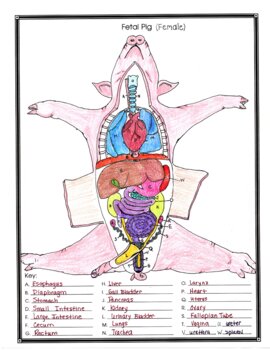
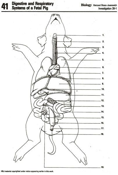
Female Fetal Pig Anatomy Dissection Guide and Worksheet

Preparation for Dissection

Before embarking on the detailed exploration of fetal pig anatomy, it’s crucial to prepare adequately. This preparation includes gathering necessary tools and ensuring your workspace is suitable for the dissection.
- Tools Needed:
- Scalpel or sharp scissors
- Dissecting pins
- Dissecting tray
- Forceps
- Probes
- Dissection manual or guide
- Gloves
- Apron or lab coat
- Face mask (optional for odor sensitivity)
- Workspace Preparation:
- Clean and disinfect your workspace.
- Ensure good lighting for detailed examination.
- Have a trash bag or disposal bin for organic waste.
- Keep cleaning supplies nearby for post-dissection cleanup.
🧪 Note: Always use appropriate safety measures when performing dissections, such as wearing protective gloves and disposing of biological material properly according to local regulations.
External Anatomy

Understanding the external features of a fetal pig can provide insights into its development and comparative anatomy with humans.
- Anatomical Landmarks: Identify and mark:
- The snout, nostrils, and mouth.
- Eyes, ears, and body orientation.
- Teats and urogenital openings to determine sex.
Examine the pig's overall external anatomy, noting any differences between the species and human anatomy:
- Head Region: Look for the umbilical cord’s remnant (a feature absent in humans).
- Body Cavities: Determine the sex by observing the genital papilla in males and the location of urogenital openings in females.
📚 Note: Referring to your guide or manual can help in identifying the less obvious features.
Internal Anatomy

Once you’ve familiarized yourself with the external anatomy, it’s time to delve into the fetal pig internal anatomy. Follow these steps for a comprehensive dissection:
Opening the Body Cavities

- Incisions: Make a series of incisions to access the internal organs. Here’s how:
- Start with an incision at the umbilical cord, moving caudally along the ventral surface towards the anus.
- Make an additional “Y” incision on the thorax to spread the ribcage.
- Extend the incision around the legs for better access to the abdominal cavity.
| Region | Incision Description |
|---|---|
| Abdominal | From umbilical cord to anal region, cut through skin and muscle layers. |
| Thoracic | A "Y" incision starting at the throat, spreading the ribcage. |
| Extremities | Cut around limbs for better exposure of the abdominal cavity. |

Examining the Thoracic Cavity

- Organs: Inspect the heart, lungs, and thymus gland.
- Identify the pericardium enveloping the heart.
- Note the left and right lungs, their lobes, and the presence of pleural membranes.
- Observe the thymus gland located near the heart.
Exploring the Abdominal Cavity

- Organs: Carefully examine the organs in this region.
- Liver: Note its four lobes.
- Gallbladder: Examine its connection to the liver.
- Stomach: Observe the cardiac and pyloric sphincters.
- Intestines: Differentiate the small intestine from the large intestine.
- Spleen, pancreas, and kidneys: Identify their positions and relationships with other organs.
📝 Note: Be gentle when handling internal organs to prevent damage. Use pins to keep incisions open or to secure tissues.
Examining the Female Reproductive System

- Organs: Here’s what to look for:
- Ovaries and Oviducts: Trace their path from the horns of the uterus.
- Uterus: Note its “Y” shape, with the uterus horns leading to the cervix.
- Vagina and Urethra: Observe their junction and the urogenital sinus.
Cleanup and Disposal

After completing the dissection, ensure you follow proper cleanup and disposal procedures:
- Cleanup:
- Rinse off any residual tissue from tools.
- Clean and disinfect all surfaces used.
- Dispose of gloves and other disposables.
- Disposal:
- Place organic waste in a designated biohazard bag.
- Ensure chemicals used during preservation are disposed of according to safety guidelines.
As we draw to a close on this dissection journey, it’s clear how engaging with fetal pig anatomy provides not just anatomical insights but also connects us to biological science, highlighting the complexities of life and its development. By methodically dissecting the pig, from its external features to its internal intricacies, we’ve journeyed through a fundamental experience that fosters a deep appreciation for anatomical study.
This dissection has allowed us to make comparisons with human anatomy, observe the development stages of an organism, and understand the workings of various organ systems. Moreover, the process emphasizes the importance of preparation, ethical treatment of specimens, and adherence to proper scientific protocols.
In reflecting on this exploration, we recognize the significance of biology in understanding life. Each step of the dissection was not just about cutting or removing; it was an exercise in precision, patience, and respect for the scientific process.
Why is fetal pig dissection used in educational settings?

+
Fetal pig dissection provides students with a hands-on learning experience, allowing them to understand mammalian anatomy, organ systems, and comparative physiology. It’s an effective teaching tool for biology and anatomy classes.
How do fetal pigs differ from adult pigs anatomically?

+
Fetal pigs lack many of the adult features like developed sex organs, fully formed hooves, and a more complex digestive system. Their organs are generally smaller and less differentiated than those of adult pigs.
What precautions should be taken during dissection?

+
Use protective gear like gloves and aprons. Ensure good ventilation to manage any smells, maintain a clean workspace, and handle the specimen gently to avoid damaging delicate organs. Follow proper disposal methods for biological waste.


