Synapse Worksheet: Dive into Anatomy
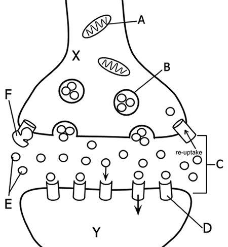
In the intricate landscape of human anatomy, every structure has a purpose and a function. Among these, the synapses stand out as the critical junctions where neurons communicate, making them central to our understanding of neurological processes. Today, we will delve deep into the world of synapses, exploring their anatomy, function, and clinical relevance.
Anatomy of a Synapse

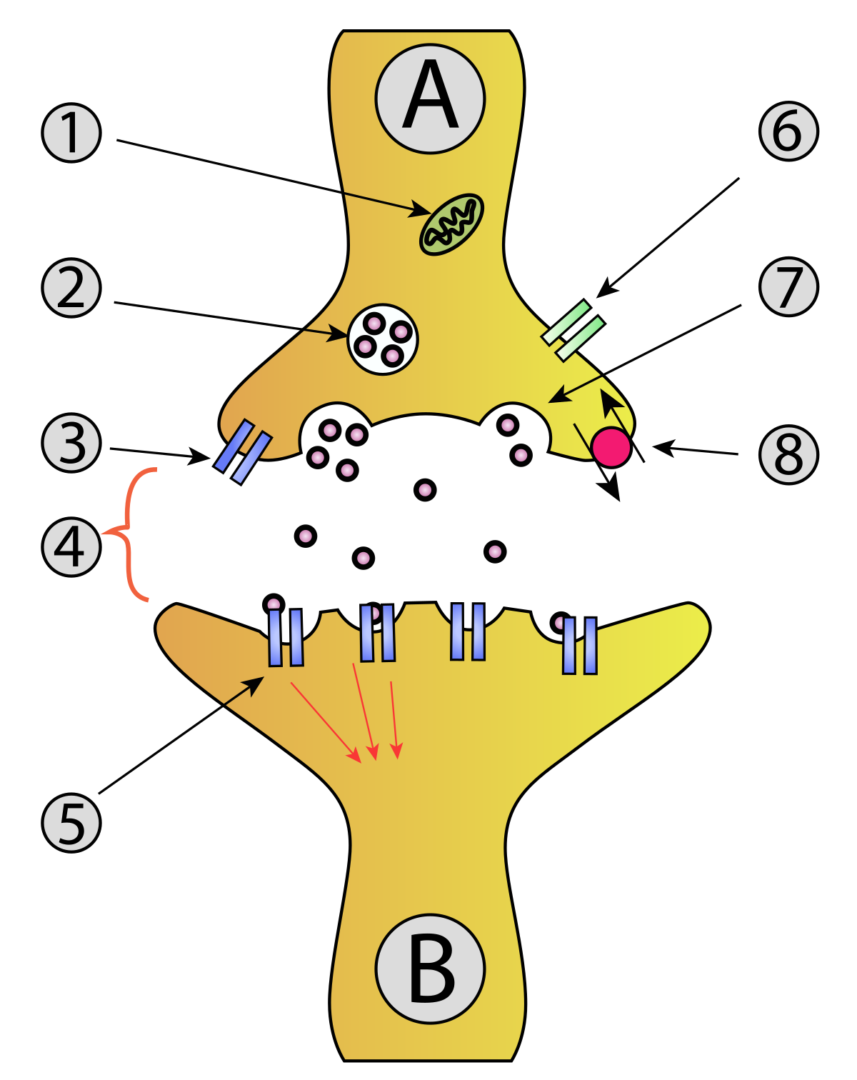
A synapse is not just a simple gap between two neurons; it is a highly organized structure designed for efficient and selective communication. Here are the key anatomical components:
- Presynaptic Neuron: This neuron sends information. It contains vesicles with neurotransmitters ready to be released into the synaptic cleft.
- Postsynaptic Neuron: Receives the signal. The postsynaptic membrane contains various receptors to which neurotransmitters bind.
- Synaptic Cleft: The tiny gap (about 20-40 nm wide) between the pre- and postsynaptic neurons where neurotransmitters are released and travel to bind with receptors.
- Synaptic Vesicles: These are membrane-bound sacs within the presynaptic terminal, storing neurotransmitters.
- Synaptic Cleft: Not just a gap, but filled with extracellular fluid, containing adhesion molecules and enzymes that modulate synaptic activity.
Types of Synapses

Synapses can be classified based on their function or location:
- Chemical Synapses: These are the most common, where neurotransmitters act as chemical messengers to transmit signals across the synaptic cleft.
- Electrical Synapses: Here, neurons communicate directly through gap junctions, allowing for rapid, bidirectional signal transfer.
- Neuromuscular Junctions: A specialized synapse between motor neurons and muscle fibers, pivotal for movement.
- Autoreceptors: Found on the presynaptic neuron, these can detect the neurotransmitter levels in the synapse, controlling further release.
Synaptic Transmission
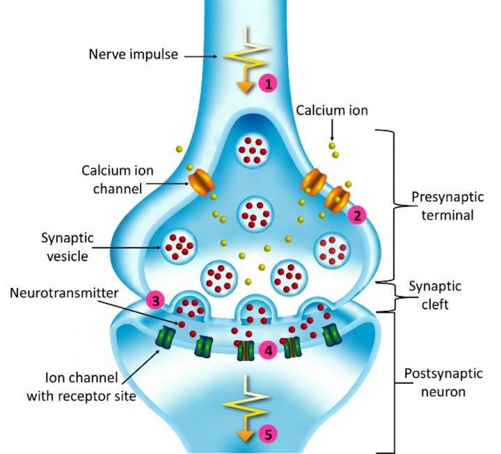
Let's break down the process:
- Action Potential Arrival: An electrical impulse (action potential) arrives at the presynaptic terminal.
- Calcium Entry: This causes voltage-gated calcium channels to open, allowing calcium ions to enter the presynaptic terminal.
- Vesicle Release: The influx of calcium causes synaptic vesicles to fuse with the presynaptic membrane, releasing neurotransmitters into the synaptic cleft via exocytosis.
- Neurotransmitter Binding: Released neurotransmitters travel across the cleft and bind to postsynaptic receptors.
- Signal Propagation: The binding either excites or inhibits the postsynaptic neuron, depending on the type of neurotransmitter and receptor.
- Signal Termination: Neurotransmitters are either degraded, reuptaken by the presynaptic neuron, or diffuse away from the synapse.
📌 Note: Each step in synaptic transmission is crucial for ensuring effective neuronal communication. A malfunction at any point can lead to neurological disorders.
Synaptic Plasticity
This term refers to the ability of synapses to strengthen or weaken over time, in response to increases or decreases in their activity. Here's what it entails:
- Long-Term Potentiation (LTP): A long-lasting enhancement in signal transmission between neurons. This is thought to underlie learning and memory.
- Long-Term Depression (LTD): Conversely, a sustained decrease in synaptic efficacy.
Clinical Relevance

Synapses are not just a topic for anatomical study; they have profound clinical implications:
| Condition | Synaptic Malfunction | Clinical Manifestation |
|---|---|---|
| Alzheimer's Disease | Disruption in neurotransmitter balance, especially acetylcholine | Memory loss, confusion, decline in cognitive functions |
| Parkinson's Disease | Depletion of dopamine at synapses in basal ganglia | Movement disorders, tremors, rigidity |
| Depression | Alteration in serotonin, norepinephrine, and dopamine levels | Mood changes, loss of interest, cognitive dysfunction |
| Epilepsy | Excessive synchronized neuronal firing due to altered synaptic inhibition | Seizures |

💡 Note: While treatments target these synaptic changes, understanding the exact mechanisms remains a frontier in neuroscience research.
To wrap up, the synapse, with its complex yet elegant structure, is the linchpin of neural communication. Our exploration highlights the intricacies of synaptic function, its pivotal role in our cognitive processes, and how its study not only enlightens us about our brain but also aids in addressing neurological disorders. The dynamic nature of synapses underscores their significance in our lives, from learning new skills to forming memories, and even in maintaining our mental health.
What are the main functions of a synapse?

+
Synapses facilitate communication between neurons by transmitting signals in the form of neurotransmitters, allowing for the integration of complex neural networks and the processing of information in the brain.
How do synapses relate to learning and memory?
+
Synaptic plasticity, which includes changes like LTP and LTD, is crucial for learning and memory. Stronger or weaker synapses can form new neural pathways or enhance existing ones, essentially rewiring the brain based on experiences and learning.
What happens when there’s a problem with synaptic function?

+
Dysfunction in synaptic transmission can lead to various neurological and psychiatric disorders. For instance, Alzheimer’s involves a loss of synapses, while depression might involve diminished neurotransmitter activity.


