5 Key Insights into Sliding Filament Theory

Understanding the mechanics behind muscle contraction can be quite an intricate process, particularly with the sliding filament theory. This theory is fundamental in physiology, offering a comprehensive explanation of how muscles work. Here, we will delve into five key insights that encapsulate the essence of this theory:
1. The Basic Concept of Muscle Contraction


The sliding filament theory, first proposed in the 1950s by Andrew Huxley and Rolf Niedergerke, along with Jean Hanson and Hugh Huxley, posits that muscle contraction occurs due to the sliding motion of actin and myosin filaments within muscle fibers. Here’s a breakdown:
- Actin: Thin filament made up of proteins called actin monomers arranged in a double helix.
- Myosin: Thick filament composed of myosin heads or cross-bridges, responsible for the power stroke.
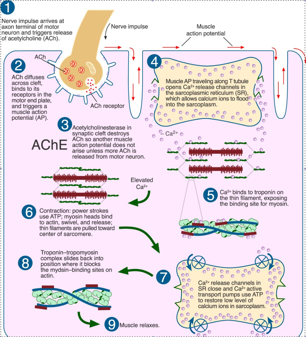
- The contraction begins when calcium ions are released into the sarcoplasm, allowing myosin to interact with actin, forming cross-bridges.
- With each ATP molecule hydrolyzed, myosin heads swivel, causing actin to slide over myosin, shortening the muscle fiber.
2. The Role of Calcium and ATP

Calcium ions play a pivotal role in initiating muscle contraction:
- Calcium ions bind to troponin, which alters tropomyosin’s position, exposing the binding sites on actin for myosin heads.
- ATP is crucial for both the detachment of myosin from actin and the repositioning of the myosin head for another power stroke.
- Without ATP, muscle relaxation is impossible as myosin remains attached to actin, leading to a condition known as rigor mortis.
3. The Interplay of Contractile Proteins


Muscle contraction involves several proteins:
- Actin: The thin filament on which myosin heads walk.
- Myosin: The motor protein that uses ATP hydrolysis for movement.
- Tropomyosin: Blocks myosin binding sites on actin in relaxed muscle.
- Troponin: Calcium-sensitive protein that regulates the position of tropomyosin.
⚠️ Note: The proteins actin, myosin, troponin, and tropomyosin work in concert to control muscle contraction.
4. Sarcomere Dynamics

| Phase of Contraction | Distance Between Z-discs | Length of A-band | I-band Length |
|---|---|---|---|
| Relaxed | Long | Constant | Long |
| Contracted | Short | Constant | Short |

The sarcomere, the basic contractile unit of a muscle fiber, shortens during contraction:
- Actin filaments slide inwards, reducing the distance between the Z-discs.
- The length of the A-band, made of myosin, remains constant.
- The I-band, composed of actin only, shortens significantly.
5. The Sarcomere’s Internal Structure

The sarcomere’s internal structure is crucial for muscle function:
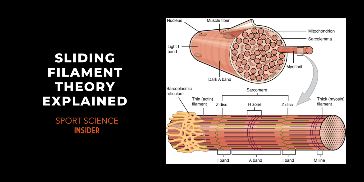
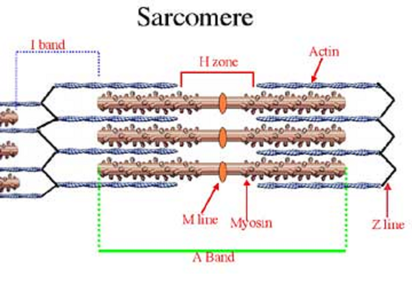
- M-line: Holds myosin at the center of the sarcomere.
- Z-discs: Anchor the ends of the actin filaments.
- H-zone: The region where only myosin is present, which shortens or disappears during contraction.
- I-band: The light band of the sarcomere, only containing actin.
To grasp the intricacies of muscle contraction, one must appreciate the dynamic interplay of these structural components. This understanding not only explains how muscles shorten, but also why muscle tone is essential for our daily activities, from the simplest to the most strenuous.
What triggers the sliding filament mechanism?

+
The binding of calcium to troponin is the trigger for the sliding filament mechanism. This binding shifts tropomyosin, exposing the actin’s binding sites for myosin interaction.
How is energy utilized in the sliding filament theory?

+
ATP is used for both the initial binding of myosin to actin and for the subsequent power stroke, where energy from ATP hydrolysis is released.
Can muscle fibers lengthen as per the sliding filament theory?

+
Yes, when myosin releases from actin and no further ATP-driven power strokes occur, the muscle fiber can lengthen passively under external forces or relax back to its resting state.