Microscope Labeling Worksheet Answers Explained: See Clearly Now

In the realm of microscopy, labeling and understanding the various components of a microscope can significantly improve one's ability to use this powerful tool effectively. Whether you're a student struggling with your first biology lab or an amateur enthusiast looking to delve deeper into the world beneath the lens, this guide will walk you through the microscope labeling worksheet answers. Not only will it help clarify the parts of the microscope, but it will also enhance your ability to utilize it for educational and scientific purposes.
Parts of the Compound Microscope

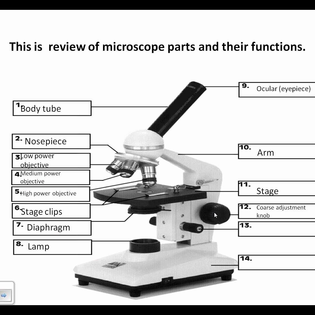
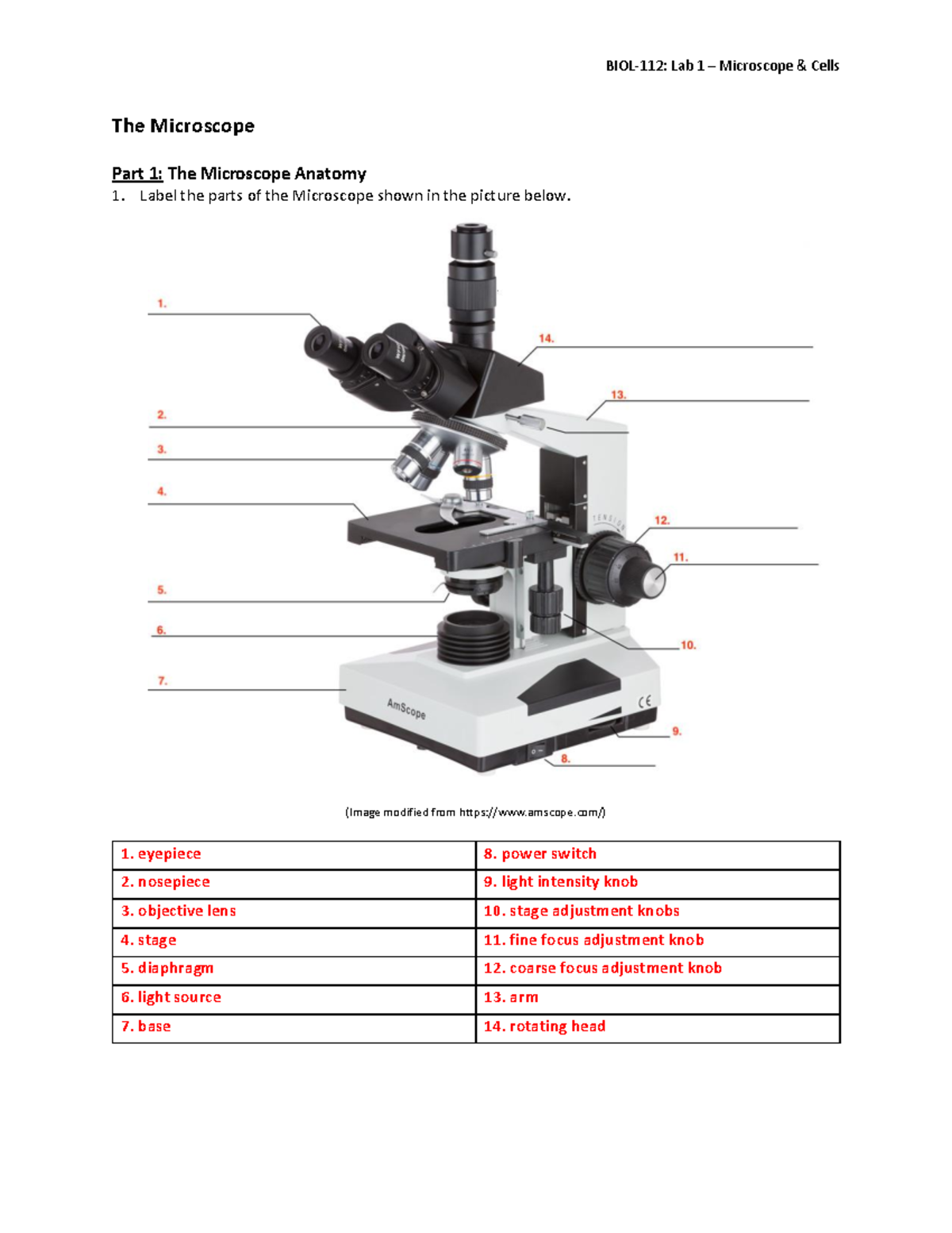
A compound microscope, the most commonly used in educational settings, has several parts, each with a specific function:
- Eyepiece: This is where the magic begins. The eyepiece, often with 10x magnification, is where you look through to view your specimen.
- Ocular Lens: Part of the eyepiece, responsible for the initial magnification of the image.
- Objective Lenses: Typically, you have three or four of these under the stage. They range from 4x to 100x in magnification, each suited for different magnifications and tasks.
- Revolving Nosepiece: This allows you to switch between different objective lenses easily.
- Stage: Where the specimen slide is placed.
- Stage Clips: Hold the slide in place.
- Condenser: Focuses light onto the specimen.
- Diaphragm: Controls the amount of light that passes through the condenser.
- Coarse Adjustment Knob: Moves the stage up or down quickly for focusing.
- Fine Adjustment Knob: Fine-tunes focus at higher magnifications.
- Body Tube: Holds the eyepiece and objective lenses at the correct working distance.
- Arm: You support the microscope with this, and it contains many internal components.
- Base: Provides stability to the entire unit.
Common Questions on Microscope Labeling

Understanding how to label a microscope diagram correctly is fundamental:
- How to Label the Eyepiece and Objective Lenses? - Label the eyepiece as the “eyepiece,” and each objective lens with its magnification power, e.g., “4x Objective.”
- Why Label the Revolving Nosepiece? - Because it’s where the objectives are mounted, allowing easy transitions between magnifications.
- What’s the Role of the Condenser? - It focuses light onto the specimen to improve image clarity.
How to Use a Microscope Labeling Worksheet

Using a microscope labeling worksheet is straightforward, but here are some tips to maximize your learning:
- Identify Parts: First, familiarize yourself with the names and functions of each part. Use your worksheet to label the microscope diagram as you go.
- Practice: Label the microscope repeatedly until you can do it by heart.
- Understand: Go beyond labeling. Know why each part is important and how it contributes to the imaging process.
- Check Accuracy: Use keys or answer guides to ensure your labeling is correct. Remember, precision in biology is key.
- Apply: After labeling, use the microscope to view slides. This practical application will solidify your understanding of each part.
💡 Note: Remember, the placement of labels should be clear to ensure readability.
Common Mistakes in Microscope Labeling

Here are some frequent errors to avoid:
- Confusing the Condenser with the Diaphragm: These are different, with the condenser focusing light and the diaphragm controlling its amount.
- Mislabeling the Arm and Base: They are crucial for microscope stability and support but often get mixed up.
- Overlooking the Fine Adjustment Knob: Especially at higher magnifications, this knob is essential for clear focus.
📝 Note: Proper labeling will help you understand how to use the microscope correctly for different specimens.
Conclusion

Understanding the intricacies of microscope labeling worksheets is not just an academic exercise; it’s a gateway to a clearer view of microscopic worlds. From the eyepiece where we begin our visual journey to the base that holds everything steady, each component plays a critical role in the magnification and examination process. By thoroughly engaging with these worksheets, you’ll not only master the terminology but also enhance your ability to use the microscope to its full potential. With practice, the once complex array of parts will become familiar tools, enabling you to explore and understand the minute details of the biological world with precision and wonder.
Why is it important to know the parts of a microscope?

+
Knowing the parts of a microscope allows you to correctly assemble, use, and maintain the instrument. Understanding each part’s function enhances your ability to troubleshoot issues and optimize imaging.
Can I change the magnification of my microscope?

+
Yes, you can change the magnification by rotating the revolving nosepiece to select different objective lenses, each providing a different level of magnification.
How do I ensure clear images under the microscope?

+
To ensure clear images, adjust the condenser, diaphragm, and use both the coarse and fine adjustment knobs to focus. Also, make sure the slide is well-prepared and clean.
What’s the difference between a compound and a simple microscope?

+
A simple microscope uses one lens for magnification while a compound microscope, which is more common in labs, uses two sets of lenses (objective and ocular) to achieve higher magnification and resolution.
Are there any safety precautions to keep in mind while using a microscope?

+
Yes, always use the coarse adjustment knob first to bring the specimen into focus, avoid touching the lenses with your fingers, keep the microscope clean, and when not in use, cover it to prevent dust accumulation.


