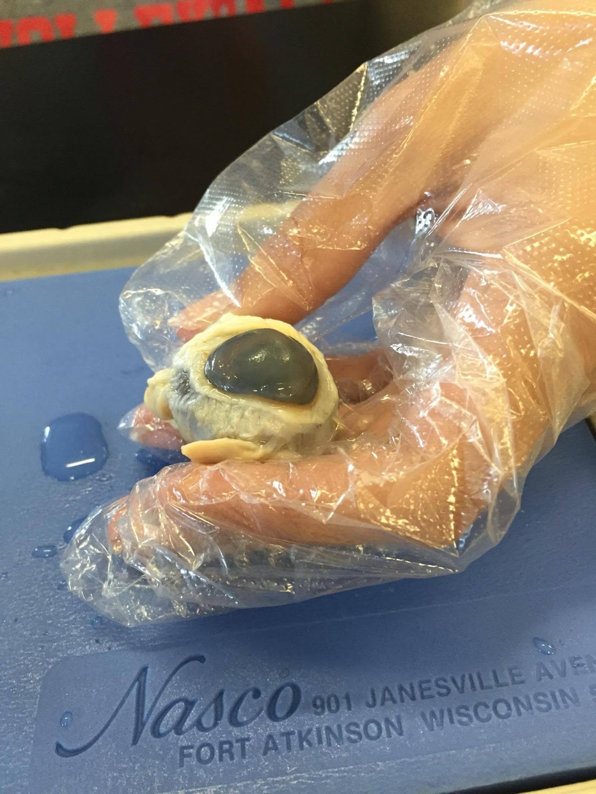
5 Steps to Dissect a Cow or Sheep Eye

Ever wondered what lies behind the visual world of animals like cows or sheep? Dissecting their eyes isn't just a peculiar educational exercise; it opens up a fascinating window into understanding biological mechanisms and anatomy. Whether you're a student preparing for a biology class or a curious mind seeking to delve into veterinary science, this guide offers a step-by-step journey through the dissection of a cow or sheep eye. Here's how you can perform this interesting dissection, ensuring you gather the most educational value while exploring the biological marvel of eye anatomy.
Preparation for Dissection


Before embarking on the dissection, preparation is key:
- Safety First: Wear gloves, protective eyewear, and an apron. This isn’t just for cleanliness but for safety.
- Secure a Specimen: Obtain a fresh or preserved cow or sheep eye from a biological supply company or a veterinary school.
- Gather Tools: You’ll need a dissection kit which includes scissors, a scalpel, a probe, and tweezers.
- Workspace Setup: Use a tray with a dissecting pad to work on, ensuring it has a drainage area for fluids.
Examine the External Features

Upon receiving your specimen:
- Identify the cornea, the clear, protective layer at the front.
- Observe the sclera, the white, outer layer of the eye.
- Locate the optic nerve, a thick cord at the back of the eye.
- Look for the extrinsic muscles, which control eye movement.
Cutting and Internal Examination

Now, let’s get inside:
- Corneal Incision: With a sharp scalpel, make a careful cut on the cornea towards the sclera. Circular movements work best to cut around the circumference of the eye.
- Remove the Lens: After lifting the cornea, locate and gently remove the clear, jelly-like lens.
- Vitreous Humor: The vitreous humor, the jelly-like substance filling the eye’s back chamber, might spill out.
Dissection of the Posterior Chamber

To explore further:
- Retina Examination: Carefully lift the optic nerve and inspect the retina, noting its color, texture, and delicate structure.
- Choroid and Tapetum Lucidum: Look at the choroid, which contains blood vessels, and in some animals, the reflective tapetum lucidum.
- Ciliary Body: Identify the ciliary body, responsible for controlling the shape of the lens.
Cleaning and Observation

After dissection:
- Rinse the remaining eye structures to remove residual fluids or tissues.
- Observe and note the aqueous humor in the anterior chamber, if any.
- Compare and contrast the iris and pupil’s size and shape with human eyes.
📝 Note: Ensure all tools are sterilized or replaced after use to prevent contamination or cross-infection.
Understanding the eye through dissection not only provides insights into how animals perceive the world but also underscores the similarities and differences with human anatomy. Each step of the process, from preparation to meticulous observation, enhances your appreciation for biological structures and their functions.
Can I use any animal eye for dissection?

+
While cow or sheep eyes are commonly used due to their similarity to human eyes, pig eyes are also viable. However, ensure the eyes are fresh or preserved correctly for the best educational experience.
What can I learn from dissecting an animal eye?

+
You can learn about the structure of the eye, how it functions, and compare differences and similarities with human eyes. It’s particularly useful for understanding visual acuity, color perception, and depth perception in animals.
How can I ensure a clean and safe dissection process?

+
Follow safety protocols by wearing protective gear, using sterilized tools, and properly disposing of biological waste. Always work in a well-ventilated area to mitigate odor and ensure safe handling of the specimen.