Axial Skeleton Labeling Worksheet Guide: Master Anatomy Easily

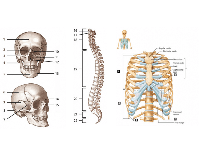
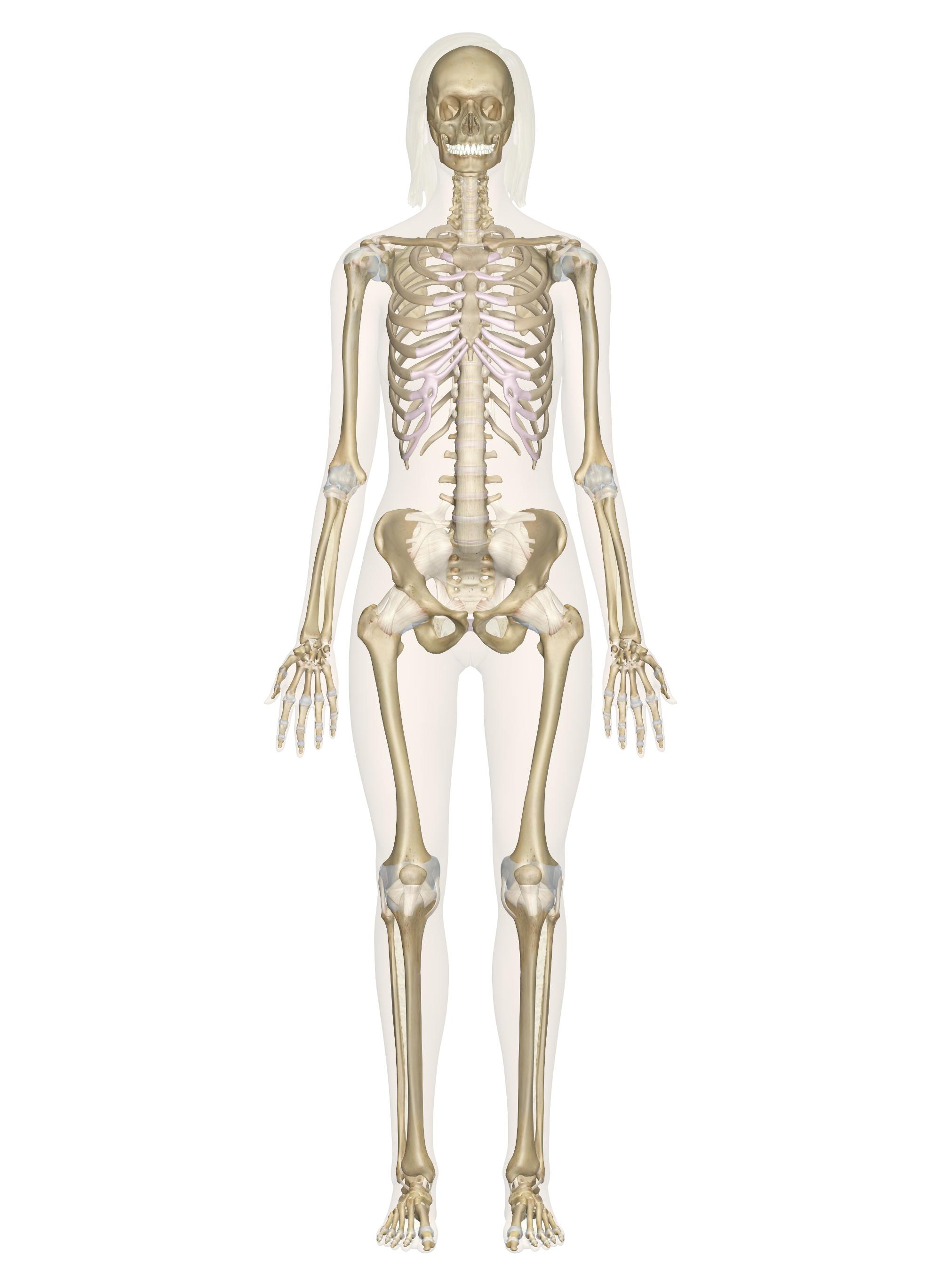
Understanding the human skeletal system can be an overwhelming task, especially when it comes to memorizing the names, positions, and functions of each bone within the axial skeleton. The axial skeleton, which consists of the skull, vertebral column, rib cage, and hyoid bone, serves as the core framework for our body, providing support, stability, and protection for vital organs. This guide offers a step-by-step approach to mastering the axial skeleton through the effective use of labeling worksheets. By engaging with these tools, you can enhance your knowledge of anatomy in a way that is both fun and educational.
The Importance of Labeling Worksheets

Labeling worksheets are more than just exercises in naming parts. They are:
- Visual Learning Tools: They cater to visual learners by providing a clear layout of anatomical structures.
- Memory Aids: Writing down names repeatedly helps in memorizing bone names and their placement.
- Engagement Boosters: Active engagement with the material through labeling can make learning anatomy less tedious.
⚠️ Note: Always ensure the worksheets are up-to-date and accurate to reflect current anatomical knowledge.
Getting Started with Axial Skeleton Labeling

Gather Your Materials

To begin labeling the axial skeleton:
- Acquire or download axial skeleton diagrams from reliable anatomy sources.
- Use colored pencils or markers to differentiate different sections.
- Have a pen or pencil for writing labels.
- Ensure you have access to an anatomy textbook or a reliable online resource for reference.
How to Label the Axial Skeleton

Follow these steps to effectively label your axial skeleton worksheet:
- Identify the Sections: Start by recognizing the main parts: skull, vertebral column, rib cage, and hyoid bone.
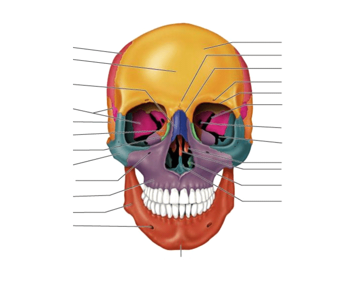
- Locate Specific Bones: Within each section, identify individual bones. For example, within the skull, locate the frontal, parietal, temporal, occipital, and other bones.
- Write Labels: Carefully write the name of each bone in its corresponding location, ensuring your handwriting is neat for future reference.
- Color Coding: Use different colors to mark different bone groups, which will help in memorization through visual cues.
- Check and Correct: Use your reference material to verify your labels. Correct any mistakes and add any bones you missed.
| Bone | Section | Function |
|---|---|---|
| Frontal Bone | Skull | Forms the forehead and roof of the eye sockets |
| Vertebrae | Vertebral Column | Support and protect the spinal cord |
| Ribs | Rib Cage | Protect internal organs and assist in breathing |

Tips for Effective Learning

- Repetition: Repeat the labeling process several times, as repetition aids memory retention.
- Quiz Yourself: Create quizzes where you label without looking at your reference. This helps assess your knowledge and reinforce learning.
- Mnemonics: Use mnemonics to remember bone names or their functions. For instance, “FATTOM” for the skull bones: Frontal, Axis (C1), Temporal, Temporal, Occipital, and Mastoid (part of the temporal bone).
- Practice with Different Angles: Try labeling from different perspectives to understand the 3D arrangement of bones.
- Study Groups: Learning with peers can provide different perspectives and help in explaining concepts to each other.
💡 Note: Use digital tools like anatomy apps or interactive websites for dynamic labeling experiences.
Integrating Labeling with Other Learning Methods

Labeling alone is beneficial, but it should be part of a broader study strategy:
- Dissection or 3D Models: Combine your worksheet learning with practical experience if possible.
- Anatomy Texts: Refer to comprehensive textbooks for in-depth details about each bone’s function and structure.
- Online Resources: Utilize online interactive anatomy programs for visual aids and self-testing.
- Videos: Educational videos can provide a dynamic view of the skeletal system in motion.
By integrating labeling with these methods, you can gain a more thorough understanding of the axial skeleton.
Mastering the axial skeleton through labeling worksheets is not just about memorization but also about understanding the framework of the human body. This approach allows for an interactive and engaging learning process, catering to different learning styles and ensuring that the knowledge sticks. By following these steps, using tips for effective learning, and integrating labeling with other methods, you can achieve a comprehensive grasp of the axial skeleton's anatomy. Whether for academic purposes or personal interest, this guide aims to make the journey through anatomy not only educational but also enjoyable.
What are the primary components of the axial skeleton?

+
The primary components are the skull, vertebral column, rib cage, and hyoid bone.
Why use coloring in anatomy labeling?

+
Coloring helps in visual differentiation of bone groups, enhancing memory through visual association.
How often should I practice labeling?

+
Frequent practice, ideally several times a week, helps with retention and recall.
Can this method help with exams?

+
Yes, by practicing labeling, you not only memorize but also understand the spatial relationships of bones, which can be crucial during anatomy exams.